Non-invasive, laser doppler system detects skin cancer
Source: optics.com. August 2015
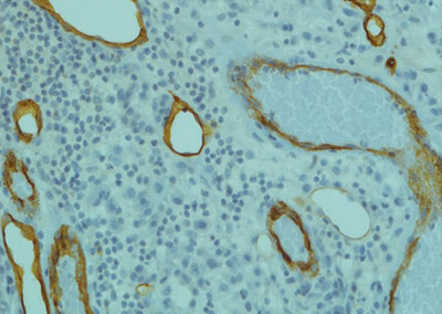
Skin malignant melanoma is a highly angiogenic cancer – it develops via blood vessels – necessitating early diagnosis for positive prognosis. But the current diagnostic standard of biopsy and histological examination inevitably leads to many unnecessary invasive excisions.

Source:Optics.com
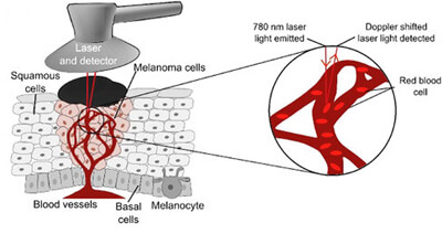
Now, researchers at Lancaster University, UK, in partnership with Pisa University, Italy, have developed a new non-invasive technique which can accurately detect malignant melanoma without a biopsy.
In particular, the team led by Professor Aneta Stefanovska looks at the results from scanning a sample across the frequency range 0.005–2Hz, which is associated with both local vascular regulation and effects of cardiac pulsation. The work was recently published in Nature Scientific Reports.
The paper reports that the laser technique to detect the subtle differences in blood flow beneath the skin enables researchers to tell the difference between malignant melanoma and non-cancerous moles. During the study, 55 patients with atypical moles agreed to have their skin monitored by researchers at Pisa University Hospital using a laser Doppler system. The laser Doppler recorded the complex interactions taking place in the minute blood vessels beneath their suspicious mole for around 30 minutes.
The fluctuations in recorded signals were then analyzed using methods developed by physicists at Lancaster University. The patients in the study then went on to have their moles biopsied and the results were compared with the information obtained – noninvasively – using the laser Doppler scan.
100% accuracy

Source: Optics.com
The laser Doppler signal correctly identified 100% of the patients with malignant skin. Professor Stefanovska said,
“We used our knowledge of blood flow dynamics to pick up on markers which were consistently different in the blood vessels supplying malignant moles and those beneath normal skin. Combining the new dynamical biomarkers we created a test which, based on the number of subjects tested to date, has 100% sensitivity and 90.9% specificity, which means that melanoma is identified in all cases where it is present, and ruled out in 90.9% of cases where it is not.”
Professor Marco Rossi of Pisa University added,
“Skin malignant melanoma is a particularly aggressive cancer associated with quick blood vessel growth which means early diagnosis is vital for a good prognosis. The current diagnostic tools of examination by doctors followed by biopsy inevitably leads to many unnecessary invasive excisions. This simple, accurate, in vivo distinction between malignant melanoma and atypical moles may lead to a substantial reduction in the number of biopsies currently undertaken"
Next steps
The Nature Scientific Reports paper concludes, “To translate these results to a larger scale, and facilitate the development of a specialized ‘melanometer’, the inherent heterogeneity of melanoma and atypical nevi lesions necessitates the recruitment of a larger cohort, as part of a multi-centre study. This should incorporate a wider range of lesion subtypes, for example Spitz nevi and melanoma in situ, to verify applicability of the results to all diagnostically difficult pathologies.

Source: Optics.com
“A multi-channel Laser Doppler Flowmetry system can be developed for this specific purpose, and the knowledge that the lowest frequency interval of interest is 0.02?Hz will allow a reduction in measurement time to only 15min. If successfully verified, this method is fast, inexpensive and non-subjective, and requires minimal training, providing huge potential for clinical use, and even accessibility to the public in the form of a specialized device for monitoring the evolution of skin lesions.”
