Antigen processing in melanoma
Dr Katherine Woods
T cells can recognize and kill melanoma cells by targeting short pieces of protein on the melanoma cell surface, called epitopes. The epitopes are made inside the melanoma cell where proteins are chopped into small pieces in a complex called the proteasome.

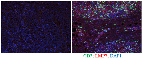
Depending on inflammation at the tumour site, the proteasome can change the way it chops the proteins, and therefore which epitopes are presented on the cell surface. In this way the tumour can escape from immune cell killing, since the T cells cannot recognize and target the changed epitopes. We want to see whether the type of proteasome expressed by a tumour can predict whether a patient is more likely to successfully respond to therapy. We will use human biopsy samples and stain them with a new technique where several different targets can be identified by multicolour staining (Figure 1). In doing this study we hope to develop a method that will aid in the selection of the best treatment option for any given patient.
1= Woods K, Knights AJ, Anaka M, Schittenhelm RB, Purcell AW, Behren A, Cebon J. Mismatch in epitope specificities between IFN? inflamed and uninflamed conditions leads to escape from T lymphocyte killing in melanoma. J Immunother Cancer. 2016 Feb 16;4:10
